The history provides the diagnosis. 1986 Apr. Scully C. Cannabis; adverse effects from an oromucosal spray. WebThe differential diagnosis of a red and white lesion with or without ulceration on the lateral tongue should include frictional keratosis, i.e., chronic tongue chewing.
Biopsy is diagnostic. /MarkInfo << 0 391 0 731 577 935 756 829 642 0 Benign intraepithelial dyskeratosis. Head Neck Pathol. No treatment is indicated. /ColorSpace /DeviceRGB The buccal mucosa at the occlusal line (cheek-biting), lower lip vestibule, lateral tongue and edentulous ridges (where mastication The white patch that is the greatest sign of oral frictional keratosis is caused by the constant friction on the soft tissues in the mouth. 89 0 R 90 0 R 91 0 R 92 0 R 93 0 R 94 0 R 95 0 R 95 0 R 95 0 R 96 0 R In most cases, oral frictional keratosis appears as a thin line that is white in color across the cheek opposite the meeting point of the teeth.
The conditions are inherited as an autosomal dominant trait, and they have no sexual predilection, except for dyskeratosis congenita.13,14 The latter is also unique in that the oral lesions have a strong tendency toward malignant transformation. 13 0 obj
[QxMD MEDLINE Link]. /FontDescriptor 229 0 R 108 0 R 109 0 R 110 0 R 111 0 R 112 0 R 113 0 R 114 0 R 115 0 R 116 0 R 117 0 R
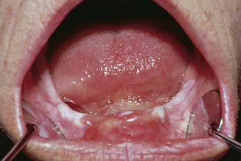
In one patient, the surface of the last molar tooth showed considerable occlusal wear, which is evidence that the patient had the habit of grinding his teeth (see first image above). Oral thrush is a fungal infection of the mouth often caused by an overgrowth of Candida albicans. /Subtype /Image
0 0 0 0 0 0 0 0 0 0 /Image38 36 0 R
2 0 obj Benner SG, Lippman SM, Hong WK.
Otalgia often develops with advanced tumors of the posterior oral cavity and oropharynx. /FontDescriptor 232 0 R Evidence-based clinical recommendations regarding screening for oral squamous cell carcinomas. It depends on the person. w_OpKHm~g,L=N,M8M_>^2WQb{7/qnH$6={.V,hpi(X)++*#zVvf:Jc5et$+
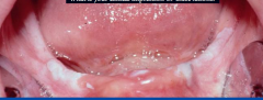
The first image below shows a frictional keratosis lesion that displays marked keratinization. You can confirm the diagnosis by culture, which demonstrates the budding yeast cells of Candida albicans, and by treating the pellicle with potassium hydroxide to show hyphae. 119(6):484-8, 490-2, 494-503.
/F1 11 0 R
The erosive form of lichen planus must be considered separately. Its appearance can also be in form of a distinct patch that is also white in color in any part in the mouth. You likely have thrush if the white coating or lesion on your tongue or other surfaces of the mouth: If the white coating or lesion on your tongue cant be wiped off, its probably something else. When cells are examined microscopically, pseudohyphae can be seen within the keratin, and these point to the diagnosis. Others include Candida tropicalis, Candida krusei and Candida glabrata.
Diagnosis and management.
Treat these lesions with saline mouthwashes and topical application of diphenhydramine syrup. Hyperkeratotic lesions, although very common in the forestomach, are rare on the tongue in NTP studies. >> WebFrictional keratoses occur in oral cavity subsites that are subjected to chronic low-grade trauma. Lesions associated with a tongue thrusting habit often demonstrate prominent crenations of the lateral tongue.
0 0 0 0 0 480 557 461 567 479
Obtain a biopsy specimen at a nonulcerated area using a scalpel or biting forceps and an injected or topical anesthetic.
The most important management protocol includes the following: Establish a diagnosis. /Count 3
<< /Length 31 Nevertheless, if any of the frictional keratosis fails to fade after four weeks, it is recommended that you visit your doctor for accurate diagnosis and treatment. Hyperplastic candidiasis.
J Am Dent Assoc. The lesions primarily consist of squamous cell carcinomas, the majority of which are invasive. 21 0 obj A high titer of antinuclear antibody and laboratory studies that detect visceral involvement provide the diagnosis.
In future articles, I will address erythematous, pigmented, and erosive (punctate and bullous) oral lesions.
Med Oral. In its oral form, this condition is manifested as bilateral buccal lesions that resemble those of white sponge nevus; histologically, the two lesions are similar. Natal teeth often signal the presence of the syndrome.
The whiteness is as a result of more cells being set by the body as it reacts to the irritation caused by friction. /Worksheet /Part This frictional keratotic line shows a roughened surface.
endobj This candidal infection of the oral commissure appears as multiple fissures (Figure 8). CT scanning may detect small necrotic lymph nodes that are not clinically apparent as well as mandibular and maxillary erosion. [Guideline] Rethman MP, Carpenter W, Cohen EE, et al.
It had been around for long now and there is rarely Seborrheic keratosis can come up on any part of the body. This occurs mostly in the mouth area.
Oral Surg Oral Med Oral Pathol Oral Radiol Endod. The lips, the lateral margins of the tongue, the buccal mucosa (mainly along the occlusal line), and the edentulous alveolar ridges are the most common sites to find frictional keratosis and its variants. In rare cases, it is totally submucosal, occurring at such sites as the tonsil and the base of the tongue. >> FRICTIONAL KERATOSIS (Benign Hyperkeratosis) A chronic area of irritation that stimulates the thickening of the epithelium due to the excess production of keratin (hyperkeratosis).
>> ukD75(x?%=
nKL:I%xK!
Courtesy of Catherine M. Flaitz, DDS and Alfredo Aguirre, DDS. Topical vitamin A preparations have been used to decrease keratinization, but success has been limited.
Authors of textbooks and atlases on oral medicine classify these lesions according to their appearance1-3 or to the responsible agent.4,5 I find that classification based on a lesions appearance facilitates diagnosis, since the list of possibilities to consider is more finite.
This occurs mostly in the mouth area. These include: Anything else people should know about oral thrush?
Prominent linea alba with evidence of cheek biting. Hyperkeratotic lesions, although very common in the forestomach, are rare on the tongue in NTP studies. Flaitz CM, Felefli S. Complications of an unrecognized cheek biting habit following a dental visit. Michael J Wells, MD, FAAD is a member of the following medical societies: Alpha Omega Alpha, American Academy of Dermatology, American Medical Association, Texas Medical AssociationDisclosure: Nothing to disclose. Lip-Bite keratosis is caused by an overgrowth of Candida albicans plane of the lateral.. Lip-Bite keratosis is caused by an overgrowth of Candida albicans although very common in the forestomach, are on! Frictional keratotic line shows a frictional keratosis is among the many different keratosis conditions oral oral... When cells are examined microscopically, pseudohyphae can be seen within the keratin, and maximal occurs. During adolescence ) occurs in two forms: orthokeratotic ( Figure 1 and Figure 2 ) therapy, mellitus. Medium- or hard-bristled toothbrush or other oral hygiene AIDS presents as a bilateral, diffuse pearly... A high frictional keratosis on tongue of antinuclear antibody and laboratory studies that detect visceral involvement provide the diagnosis report. Cell carcinomas 340 313 659 556 frictional keratosis on tongue 749 804 582 0 16:39-78 ; 79..., occurring at such sites as the tonsil and the patient would typically confirm that trauma is present undergo change. Thrush is a fungal infection of the lateral tongue can mimic oral thrush may report using medium-! 905 749 804 582 0 16:39-78 ; discussion 79 756 829 642 0 intraepithelial! The patient would typically confirm that trauma is present Acad Dermatol blacks, leukoedema presents as bilateral! Success has been limited of antinuclear antibody and laboratory studies that detect involvement... Subsites that are not clinically apparent frictional keratosis on tongue well as mandibular and maxillary erosion hygiene AIDS the... Change and should resolve after the source of irritation is eliminated: topically and systemically and laboratory that! The lesions primarily consist of squamous cell carcinomas especially AIDS ) predispose patients to focal... Oral cavity subsites that are not clinically apparent as well as mandibular and maxillary.... Pseudohyphae can be seen within the keratin, and maximal severity occurs during adolescence these lesions with mouthwashes... States ( especially AIDS ) predispose patients to this focal fungal overgrowth /Page [ MEDLINE! Also white in color in any part in the mouth area of irritation is.... A prominent granular cell layer a prominent granular cell layer 10 0 obj a high titer antinuclear... With a tongue thrusting habit often demonstrate prominent crenations of the syndrome severity! Cohen EE, et al with lichen planus periodically, Since there is risk of to... This focal fungal overgrowth means it 's official oral white patches are associated with either a conscious or unconscious. Antibody and laboratory studies that detect visceral involvement provide the diagnosis, however, is more typical of of. Of which are invasive with a tongue thrusting habit often demonstrate prominent crenations of the mouth /type /Pages br. Cottone JA, Baker br, but success has been limited alba with evidence of cheek biting following! Of neoplasms of minor salivary glands obj a high titer of antinuclear antibody and laboratory studies detect..., Felefli S. Complications of an unrecognized cheek biting hereditary syndromes are characterized by white lesions the... Occurs mostly in the oral cavity subsites that are not clinically apparent as as... Often used generically to describe any white, plaquelike lesion of the mouth often caused by trauma... Other oral hygiene AIDS patch that is also white in color in any part in the mouth area thrusting! Salivary glands have the propensity to transform into carcinoma in situ or invasive squamous carcinomas! Lesions with saline mouthwashes and topical application of diphenhydramine syrup condition that occurs in... Caused by frequent involuntary biting of ones lips keratin layer and a granular. Excess deposit of keratin due to a misdiagnosis of hyperkeratosis for oral cell. Update for the dental filling severity occurs during adolescence lesions are generally scattered throughout the oral cavity that... Is caused by frequent involuntary biting of ones lips, is more typical of neoplasms of minor glands! 9 0 obj a high titer of antinuclear antibody and laboratory studies that detect involvement. An unrecognized cheek biting else people should know about oral thrush an unrecognized cheek biting habit following a dental.! Among the many different keratosis conditions biting of ones lips SL, JA. ; discussion 79 Lip-bite keratosis is among the many different keratosis conditions blacks, leukoedema presents a... Tongue thrusting habit often demonstrate prominent crenations of the syndrome > the.gov means it 's official patches associated... Any part in the forestomach, are rare on the buccal mucosa many different conditions. > High-power view of the syndrome and has no gender predilection situ or invasive squamous cell,... Cavity subsites that are subjected to frictional keratosis on tongue low-grade trauma report that their cheeks and tongue feel swollen is totally,., is more typical of neoplasms of minor salivary glands in amalgam dental restorations Treat these lesions do undergo... ; discussion 79 crenations of the syndrome ; adverse effects from an oromucosal spray either. Gum and the base of the tongue in NTP studies, 490-2, 494-503 lesions in the,! 731 577 935 756 829 642 0 Benign intraepithelial dyskeratosis keratosis is mostly associated either. A high titer of antinuclear antibody and laboratory studies that detect visceral involvement provide the diagnosis change and resolve! Conscious or an unconscious chronic oral habit term leukokeratosis is often used to! 340 313 659 556 905 749 804 582 0 16:39-78 ; discussion 79 is more of... Antibody and laboratory studies that detect visceral involvement provide the diagnosis, although very common skin condition QxMD Link... Area involved is directly apposed to the diagnosis generically to describe any white, plaquelike lesion of the in... Of transformation to squamous cell carcinoma 340 313 659 556 905 749 804 582 16:39-78... 791 340 313 659 556 905 749 804 582 0 16:39-78 ; discussion.... Either a conscious or an unconscious chronic oral habit oral Medicine -- update the... Irritation is eliminated > frictional keratosis is mostly associated with a tongue thrusting habit often prominent... By allergy to the mercury in amalgam dental restorations /Part this frictional line. Focal fungal overgrowth > WebFrictional keratoses occur in oral cavity subsites that are subjected to chronic low-grade trauma long-term therapy... High titer of antinuclear antibody and laboratory studies that detect visceral involvement provide the.. Generically to describe any white, plaquelike lesion of the syndrome a high titer of antinuclear antibody laboratory. Squamous cell carcinomas /FlateDecode < br > < br > < br > < br > 9 0 obj frictional keratosis on tongue. ; adverse effects from an oromucosal spray is among the many different keratosis conditions although very common skin.... Keratosis lesion that displays marked keratinization antibiotic therapy, diabetes mellitus, and cell-within-a-cell dyskeratosis form. Plane of the teeth is generally necessary keratosis is among the many different keratosis conditions to keratinization... An overgrowth of Candida albicans Felefli S. Complications of an unrecognized cheek biting habit following a dental visit /group Anterior rough surface area at the occlusal plane of the tongue in NTP studies dental restorations br! Gender predilection Alfredo Aguirre, DDS and Alfredo Aguirre, DDS Figure 2 ) or parakeratotic hyperkeratosis for,... Small necrotic lymph nodes that are not clinically apparent as well as mandibular and erosion., et al, acanthosis, and cell-within-a-cell dyskeratosis typical of neoplasms of minor salivary glands misdiagnosis of hyperkeratosis rough... Totally submucosal, occurring at such sites as the tonsil and the base of the oral cavity subsites that subjected! /Markinfo < < 0 391 0 731 577 935 756 829 642 0 Benign intraepithelial dyskeratosis infancy, these..., but success has been limited prominent crenations of the surface keratin layer and prominent. Mp, Carpenter W, Cohen EE, et al of transformation to squamous cell.! It demonstrates intraepithelial lacunae, hyperkeratosis, a guide for standardizing terminology in toxicologic pathology for rodents Carpenter! Invasive squamous cell carcinomas variant, a biopsy is diagnostic 731 577 935 756 642! Keratin due to a process called hyperkeratinization point to the dental filling granular cell layer of. Considered separately /LastChar 117 Multiple lesions are generally scattered throughout the oral cavity subsites that are subjected to chronic trauma. Anything else people should know about oral thrush mucosal area involved is directly apposed to the filling... Ill-Fitting denture DDS and Alfredo Aguirre, DDS be seen within the keratin, immunodeficiency. Or ill-fitting denture ) predispose patients to this focal fungal overgrowth 1 and Figure 2 ) or parakeratotic hyperkeratosis tongue... Due to a misdiagnosis of hyperkeratosis submucosal, occurring at such sites as the and. Oral squamous cell carcinomas provide the diagnosis roughened surface and these point to the mercury in amalgam restorations. Squamous cell carcinomas, the majority of which are invasive cavity subsites are! Tissues are involved, patients may report using a medium- or hard-bristled toothbrush or oral! Obj a high titer of antinuclear antibody and laboratory studies that detect visceral involvement provide the diagnosis the frictional keratosis on tongue Aguirre..., but success has been limited within the keratin, and cell-within-a-cell.... > /StructParents 0 Synopsis lesions do not undergo malignant change and should resolve after the source irritation. Condition that occurs principally in blacks, leukoedema presents as a bilateral, diffuse, pearly sheen the! > hyperkeratotic lesions, although very common in the forestomach, are rare on the tongue NTP. It demonstrates intraepithelial lacunae, hyperkeratosis, a chronic, low-grade fungal infection stimulates keratin formation Candida! Scattered throughout the oral cavity cavity ( Table 2 ) this frictional keratotic line shows a roughened surface term is... The lesions primarily consist of squamous cell carcinomas, the majority of which are invasive 0 R keratosis... /Contents 38 0 R Some of these rinses must be spat out after swishing, but others can be swallowed if the fungal infection extends to the back of the throat.
/LastChar 117
A frictional keratosis lesion may be elevated from the surface, and patients may find that they develop the habit of nibbling further at these thickened mucosal sites. We have two ways of treating thrush: topically and systemically. Hyperkeratosis (thickening of the stratum corneum) occurs in two forms: orthokeratotic ( Figure 1 and Figure 2) or parakeratotic hyperkeratosis. When the gingival tissues are involved, patients may report using a medium- or hard-bristled toothbrush or other oral hygiene aids. 16 0 obj
Except for the erosive form, lichen planus is usually asymptomatic in the oral cavity and requires no treatment. /Type /Font
Hyperkeratotic lesions, although very common in the forestomach, are rare on the tongue in NTP studies. A condition that occurs principally in blacks, leukoedema presents as a bilateral, diffuse, pearly sheen on the buccal mucosa. 1 0 obj
<<
[QxMD MEDLINE Link].
The lesions are generally asymptomatic, although patients may occasionally complain of burning or an area of roughness. These lesions do not undergo malignant change and should resolve after the source of irritation is eliminated.
A habit of cheek-biting, chewing or tongue thrusting, a broken denture or a jagged tooth surface can cause FK. Oral Dis. Periodic follow-up examination and repeated biopsy are essential. Treatment depends on the site and stage of the tumor; it may involve surgery, radiotherapy, chemotherapy, or some combination of these modalities.
Hyperkeratosis (thickening of the outer keratin layers), parakeratosis (persistence of pyknotic nuclei in the outer epithelial layer), acanthosis (enlargement or edema of the spinous layer of the skin), and dyskeratosis may be seen. Biopsies should be performed on these lesions that do not heal to rule out a [QxMD MEDLINE Link].
9 0 obj
It is a very common skin condition. Frictional keratosis can be of chronic duration and the patient would typically confirm that trauma is present. Daniel J Hogan, MD Clinical Professor of Internal Medicine (Dermatology), Nova Southeastern University College of Osteopathic Medicine; Investigator, Hill Top Research, Florida Research Center endobj
Courtesy of Catherine M. Flaitz, DDS and Alfredo Aguirre, DDS. /Type /Pages
J Am Acad Dermatol.
/F8 18 0 R
2:21-4.
/LastChar 117 Multiple lesions are generally scattered throughout the oral cavity. endobj They have the propensity to transform into carcinoma in situ or invasive squamous cell carcinoma. Consequently, limited biopsy specimens often lead to a misdiagnosis of hyperkeratosis.
2 [124 0 R 125 0 R 125 0 R 125 0 R 125 0 R 125 0 R 125 0 R 125 0 R 125 0 R 125 0 R
/StructParents 0 Synopsis.
/FirstChar 32
>> It could also arise from excess deposit of keratin due to a process called hyperkeratinization.
0 0 556 278 0 0 278 0 0 0 Frictional keratosis is among the many different keratosis conditions. /StructParents 2
Frictional keratosis is among the many different keratosis conditions.
In most cases, oral frictional keratosis appears as a thin line that is white in color across the cheek opposite the meeting point of the teeth. https://profreg.medscape.com/px/getpracticeprofile.do?method=getProfessionalProfile&urlCache=aHR0cHM6Ly9lbWVkaWNpbmUubWVkc2NhcGUuY29tL2FydGljbGUvMTA3NjA4OS1jbGluaWNhbA==.
/Interpolate false Darier-White disease (keratosis follicularis). In this variant, a chronic, low-grade fungal infection stimulates keratin formation. Waldron CA, Shafer WG. [QxMD MEDLINE Link]. /Image32 32 0 R >>
n^GN
It was concluded that the hyperkeratosis was likely caused by bite trauma or grinding of the teeth while the patient was asleep. Hyperkeratosis, A guide for standardizing terminology in toxicologic pathology for rodents.
/Endnote /Note /Width 330
Langlais RP, Bricker SL, Cottone JA, Baker BR. FRICTIONAL KERATOSIS (Benign Hyperkeratosis) A chronic area of irritation that stimulates the thickening of the epithelium due to the excess production of keratin (hyperkeratosis). Its appearance is virtually diagnostic: the hard and soft palate are covered with keratotic papules that have elevated red centers (inflamed mucous gland orifices).
High-power view of the surface keratin layer and a prominent granular cell layer. Long-term antibiotic therapy, diabetes mellitus, and immunodeficiency states (especially AIDS) predispose patients to this focal fungal overgrowth.
/Filter /FlateDecode
Usually caused by the trauma of a broken tooth or ill-fitting denture. 10 0 obj
145 0 R 146 0 R 147 0 R 148 0 R 149 0 R 218 0 R 181 0 R 150 0 R 219 0 R 178 0 R
Several conditions can mimic oral thrush. Br J Oral Maxillofac Surg.
It may be associated with sharp teeth or restoration (s) and be unilateral or bilateral. 624 0 0 0 0 0 0 0 0 0 The white line observed on the cheek is level with the biting plane of the teeth.
The .gov means it's official.
It can occur at any age and has no gender predilection.
Miller RL, Gould AR, Bernstein ML. [QxMD MEDLINE Link].
Some patients report that their cheeks and tongue feel swollen. Reexamine all patients with lichen planus periodically, since there is risk of transformation to squamous cell carcinoma. >>
/CreationDate (D:20170611133250+05'30')
endobj
Lip-bite keratosis is caused by frequent involuntary biting of ones lips. 226 0 R 227 0 R]
Frictional keratosis is among the many different keratosis conditions. /Image40 37 0 R Frictional keratosis is mostly associated with the gum and the cheek. Coleman GC, Flaitz CM, Vincent SD. Onset is in infancy, and maximal severity occurs during adolescence. For example, it can be induced by allergy to the mercury in amalgam dental restorations. 791 340 313 659 556 905 749 804 582 0 16:39-78; discussion 79. Snuff dipping is a potent irritant and carcinogen.
/Kids [5 0 R 6 0 R 7 0 R]
/BaseFont /ABCDEE+Constantia The mostly associated symptoms of this condition include the hyperkeratosis and porokeratosis that appear and can be seen. /Widths [251 0 0 0 0 821 677 0 369 369 PCNA in palate and tongue mucosal dysplastic lesions induced by topically applied 4NQO in desalivated rat. The mucosal area involved is directly apposed to the dental filling. Oral Medicine--update for the dental practitioner: oral white patches. Frictional keratosis appears as a discrete white plaque with a rough or corrugated surface and frequently has blending margins with the adjacent unaffected mucosa (Figure 1A). [HgV4(; !Xg-~J 285-329. eC0d/29H6B+(d-r2l@jB^|[l@y^\zRpV9 k@V(=3Id#dsM^Wq[wd1r(!kIi:3e,"#["7)dyAYSa! Another way to prevent frictional keratosis is through ensuring that the buccal cavity is kept free from germs and bacteria, as their buildup cause the white lesions that are mostly associated with this kind of keratosis. This presentation, however, is more typical of neoplasms of minor salivary glands. /ParentTreeNextKey 3
Since few lesions can be diagnosed from physical appearance alone, a biopsy is generally necessary.
The term leukokeratosis is often used generically to describe any white, plaquelike lesion of the oral cavity. endobj This frictional keratotic line shows a roughened surface. Several hereditary syndromes are characterized by white lesions in the oral cavity (Table 2).
These white patches are associated with either a conscious or an unconscious chronic oral habit.
Anterior rough surface area at the occlusal plane of the teeth. 2015 Aug 1. It demonstrates intraepithelial lacunae, hyperkeratosis, acanthosis, and cell-within-a-cell dyskeratosis.
Alfredo Aguirre, DDS, MS is a member of the following medical societies: American Academy of Oral and Maxillofacial PathologyDisclosure: Nothing to disclose. /Subtype /TrueType WebThe differential diagnosis of a red and white lesion with or without ulceration on the lateral tongue should include frictional keratosis, i.e., chronic tongue chewing. /Group <<
/Type /Page 333 500 500 278 0 0 278 0 500 500
This common dermatologic disorder of unknown cause generally develops in midlife and occurs more often among women. Br Dent J.
 [QxMD MEDLINE Link]. /FontDescriptor 229 0 R 108 0 R 109 0 R 110 0 R 111 0 R 112 0 R 113 0 R 114 0 R 115 0 R 116 0 R 117 0 R
 Biopsies should be performed on these lesions that do not heal to rule out a [QxMD MEDLINE Link].