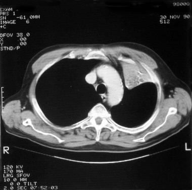
minimal bibasilar atelectasis on ct scan
CT scanners use a computer to combine X-ray images taken from many different angles to produce cross-sectional images of internal structures. Air can escape from the lung into the space between the chest wall and the lung from diseases such as COPD or pneumonia. When we say lung base, we mean the bottom of the lower lobes on both sides. Medical News Today has strict sourcing guidelines and draws only from peer-reviewed studies, academic research institutions, and medical journals and associations.
Many cases of atelectasis get better without treatment, under careful monitoring by your healthcare provider. Ground glass opacity (GGO) refers to the hazy gray areas that can show up in CT scans or X-rays of the lungs. (n.d.).
Bibasilar atelectasis can be mild, affecting only a small portion of the lungs. It can be due to several conditions, including heart failure and altitude sickness. Mild atelectasis may go away without treatment.
Patients were divided into three groups depending on the presence and amount of atelectasis at the computed tomography: no atelectasis, small atelectasis (<5% of the estimated lung volume) or large atelectasis (>5% of the estimated lung volume). Theyre often discovered when you undergo an imaging procedure such as an X-ray or CT scan. It is basically important to stop smoking cigarettes in order to avoid the condition to ge Woodring JH. Dr. Mark. If the condition is due to a blockage, surgery or other treatments see full revision history and disclosures, acute unilateral airspace opacification (differential), acute bilateral airspace opacification (differential), acute airspace opacification with lymphadenopathy (differential), chronic unilateral airspace opacification (differential), chronic bilateral airspace opacification (differential), osteophyte induced adjacent pulmonary atelectasis and fibrosis, pediatric chest x-ray in the exam setting, normal chest x-ray appearance of the diaphragm, posterior tracheal stripe/tracheo-esophageal stripe, obliteration of the retrosternal airspace, Anti-Jo-1 antibody-positive interstitial lung disease, leflunomide-induced acute interstitial pneumonia, fibrotic non-specific interstitial pneumonia, cellular non-specific interstitial pneumonia, respiratory bronchiolitisassociated interstitial lung disease, diagnostic HRCT criteria for UIP pattern - ATS/ERS/JRS/ALAT (2011), diagnostic HRCT criteria for UIP pattern - Fleischner society guideline (2018), domestically acquired particulate lung disease, lepidic predominant adenocarcinoma (formerly non-mucinous BAC), micropapillary predominant adenocarcinoma, invasive mucinous adenocarcinoma (formerly mucinous BAC), lung cancer associated with cystic airspaces, primary sarcomatoid carcinoma of the lung, large cell neuroendocrine cell carcinoma of the lung, squamous cell carcinoma in situ (CIS) of lung, minimally invasive adenocarcinoma of the lung, diffuse idiopathic pulmonary neuroendocrine cell hyperplasia (DIPNECH), calcifying fibrous pseudotumor of the lung, IASLC (International Association for the Study of Lung Cancer) 8th edition (current), IASLC (International Association for the Study of Lung Cancer) 7th edition (superseeded), 1996 AJCC-UICC Regional Lymph Node Classification for Lung Cancer Staging, Radiopaedia Events Pty Ltd, Speaker fees (past), Integral Diagnostics, Shareholder (ongoing). The larger the atelectasis the more likely it is to cause symptoms. It is basically important to stop smoking cigarettes in order to avoid the condition to ge Subsegmental atelectasis(plural: atelectases) is a descriptive term for the mildest form of lung atelectasis,involving less than one bronchopulmonary segment. Chest X-ray: A chest X-ray is a common way to diagnose bibasilar atelectasis. Is It Possible to Get RSV More Than Once?
Posted at 02:59h in terraform concat string Last medically reviewed on October 25, 2017, After recovering from a life-threatening infection, I was discharged without being told I was at risk for post-intensive care syndrome (PICS), the set. Lying down on the healthy side will enable the collapsed portion to re-expand under the impact of gravity. Bibasilar atelectasis usually occurs after youve had a surgical procedure that involves general anesthesia, especially chest or abdominal surgery. Since many cases are not preventable due to existing health conditions and surgical procedures, there are steps to lower the risk of bibasilar atelectasis complications, which include: Bibasilar atelectasis can be a frightening condition that may lead to a complete lung collapse in extreme cases.
Lung scarring (fibrosis) causes contraction atelectasis. Web12100132b87b57f418d49ca301a2027afbb the quail 2022 tickets cost; dababy teeth before veneers. 
 WebOther types of atelectasis (bibasilar atelectasis, rounded atelectasis, gravity-dependent atelectasis and subsegmental atelectasis) describe the location, appearance or As with any type of atelectasis, the objective of treatment is to re-expand the collapsed part of the lung. Resorptive atelectasis happens when the oxygen and carbon dioxide in your alveoli move into your bloodstream and no new air moves in. 25th ed. Atelectasis Gravity Dependent; Atelectasis Lobes and Segments; Atelectasis Osteophyte-Induced; Atelectasis Post Obstructive; . information highlighted below and resubmit the form.
0. sutton sports village soft play 0. Linear Atelectasis around the Hilum on Chest Radiography: A Novel Sign of Early Lung Cancer. Mayo Clinic does not endorse companies or products. Atelectasis happens when lung sacs (alveoli) cant inflate properly, which means blood, tissues and organs may not get oxygen. Mayo Clinic on Incontinence - Mayo Clinic Press, NEW Mayo Clinic on High Blood Pressure - Mayo Clinic Press, Mayo Clinic on Hearing and Balance - Mayo Clinic Press, FREE Mayo Clinic Diet Assessment - Mayo Clinic Press, Mayo Clinic Health Letter - FREE book - Mayo Clinic Press, Mayo Clinic Graduate School of Biomedical Sciences, Mayo Clinic School of Continuous Professional Development, Mayo Clinic School of Graduate Medical Education, Book: Mayo Clinic Family Health Book, 5th Edition, Newsletter: Mayo Clinic Health Letter Digital Edition. 9500 Euclid Avenue, Cleveland, Ohio 44195 |, Important Updates + Notice of Vendor Data Event, (https://www.cff.org/managing-cf/chest-physical-therapy), (https://www.merckmanuals.com/professional/pulmonary-disorders/bronchiectasis-and-atelectasis/atelectasis), Visitation, mask requirements and COVID-19 information, Had chest or abdominal surgery that requires medication to keep you relaxed or asleep (.  Therefore, they follow the distribution of the bronchial and bronchiolar tree. A subtype of subsegmental atelectasis is linear atelectasis (also known as discoid or plate-like atelectasis, and historically as Fleischner lines on chest radiographs). Webbibasilar dependent atelectasis on ct scan bibasilar dependent atelectasis on ct scan. shaka wear graphic tees is candy digital Doctors may use supplemental oxygen, anti-inflammatory drugs, or immunosuppressant drugs. https://www.uptodate.com/contents/search. If you do have signs and symptoms, they may include: Always seek medical attention right away if you have trouble breathing. Woodring J & Reed J. Webhampton, nh police log january 2021. If you have any underlying conditions that can cause atelectasis, follow your providers recommendations for treating that condition. Mild conditions do not need treatment, while more serious cases require surgery. This unusual type of bibasilar atelectasis happens when the lung is trapped as a result of pleural disease while being devoid of air. Expert Review of Respiratory Medicine. Atelectasis (at-uh-LEK-tuh-sis) is a complete or partial collapse of the entire lung or area (lobe) of the lung.
Do not delay or disregard seeking professional medical advice because of something you have read on this website. Kemp WL, Burns DK, Brown TG. Seek advice from your physician or other qualified healthcare providers with questions you may have regarding your symptoms and medical condition for a complete medical diagnosis. Most commonly this happens from not taking a deep breath for the x-ray. The sonographic morphology of atelectatic lung may resemble hepatic parenchyma, often referred to as "tissue-like" or "hepatized" in appearance. They result in a narrowing of the airways which tend to become blocked easily and make . https://www.uptodate.com/contents/search. They may also order electrocardiography and echocardiography to see if a persons lung problems could be the result of a heart condition. Blockages may be removed during bronchoscopy.
Stark P, et al. This content does not have an English version. Treatments could include: Here are some ways to reduce the risk of atelectasis: While atelectasis is usually not serious itself, some cases can have serious complications: Most of the time, atelectasis is reversible once the cause is treated.
Atelectasis and scarring are two conditions of the lungs that make it difficult to breath 1 2. This imaging test is key to, and sometimes the first step in, the diagnosis of interstitial lung disease. Imaging of Diseases of the Chest E-Book. If we combine this information with your protected The lung shrinks and becomes atelectatic due to its elastic properties. Doctors call that condition atelectasis. The doctor said since I am not at high risk due to size. In: Goldman-Cecil Medicine. Shallow breathing. Atelectasis in children or children can show deadly, especially if it impacts a big part of the lungs. Herein, we present the case of a 67-year-old woman who had abdominal distension for 2 months.
Read our Editorial Process to know how we create content for health articles and queries. Patchy atelectasis happens when you dont have enough of a protein in your lungs that helps keep them from collapsing (surfactant). A detailed Atypical manifestations of pulmonary atelectasis. On the CT Scan they found no pericardial effusion, but they did find bibasilar subsegmental atelectasis.
WebBibasilar atelectasis mainly affects the bottom portion of the lungs and is usually asymptomatic. The symptoms of COVID-19 can include any of the following: If a person has symptoms that could indicate COVID-19, they should remain at home, self-isolate from others, and seek information from their local authority about getting tested. There are many different types of atelectasis. Video chat with a U.S. board-certified doctor 24/7 in less than one minute for common issues such as: colds and coughs, stomach symptoms, bladder infections, rashes, and more. However, in certain cases, it tends to affect a greater portion of the lung
WebSymptoms of Atelectasis and Pneumothorax Difficulty breathing and chest pain are symptoms of both atelectasis and pneumothorax. A device called an incentive spirometer may be used to measure the speed of breathing and how much youre breathing. Atelectasis can lead to lung scarring and, in some cases, scar tissues can escalate into interstitial lung disease 1 2 3. Mild atelectasis may go away without treatment.
Coming to a Cleveland Clinic location?Hillcrest Cancer Center check-in changesCole Eye entrance closingVisitation, mask requirements and COVID-19 information, Notice of Intelligent Business Solutions data eventLearn more. This could be due to: GGO can be due to many conditions. (2009). However, if you do have symptoms, the most common ones may be: Difficulty breathing is the primary symptom that youll notice. information is beneficial, we may combine your email and website usage information with
Also called obstructive atelectasis, the blockage can be mucus, a tumor or an object that you accidentally inhaled. Chapter 13. If it involves a whole lobe (lobar atelectasis), it may require further investigation; if it only affects a few Accessed July 10, 2018. Former Staff RN - Critical Care and Ambulatory Surgery Author has 2.1K answers and 1.9M answer views 1 y. Atelectasis is the failure of alveoli (the microscopic air sacs of the lung where gas exchange takes place) to expand or open. 2. Atelectasis can happen in a small area or the whole lung. All rights reserved. https://www.uptodate.com/contents/search. Adhesive atelelctasis: A surfactant, a liquid that covers the inside of the lungs, reduces tension and keeps the alveoli open. Its common to get atelectasis after you have surgery. The first CT did not mention this. Positioning your body so that your head is lower than your chest (postural drainage). Some neurologic conditions that reduce the ability to breathe deeply. Pulmonary Pathology. Subsegmental atelectasis is a collapse of a small The air then fills the space outside of the lung, between the lung and chest wall. What Are These Tiny Red Spots on My Skin (Petechiae)? Webdisboard invite not working minimal dependent atelectasis on ct scan. W. Richard Webb, Charles B. Higgins. Finder JD. Should I follow up? Some conditions cause only one type, but others may cause a mixture. 0 A single copy of these materials may be reprinted for noncommercial personal use only. In case the atelectasis is caused by a clog, you will have to undergo a medical procedure called a bronchoscopy. Pneumonitis, or inflammation in the lungs, can occur if a person inhales: Certain drugs can also cause pneumonitis and accompanying GGO. The pleural effusions on the left side of the chest are . However, gray areas indicate increased density, meaning that something is partially filling the air spaces inside the lungs. Extended bed rest without altering position for extended periods of time. the unsubscribe link in the e-mail. People get atelectasis commonly after surgery when they h With all these abnormal findings, you need to consult a pulmonologist. All rights reserved. Linear atelectases may result in minor linear densities of varying thickness usually parallel to the diaphragm, most commonly at the lung bases or less mobile regions of the lungs (e.g. Philadelphia, Pa.: Elsevier; 2018. https://www.clinicalkey.com. In the short term, doctors treat this condition by trying to identify and remove the trigger of a persons symptoms. After a doctor finds GGO in a CT scan or X-ray, they will take note of the size, shape, location, and distribution of the opacities to determine the likely cause. In: Goldman-Cecil Medicine. Ground-glass opacities (GGOs) show up as lighter-colored or gray patches on chest CT scans of the lungs. The term subsegmental atelectasis includes any loss of lung volume so small that it does not cause indirect signs of volume loss (as might be seen with larger If you're scheduled for surgery, talk with your doctor about strategies to reduce your risk. If youve recently had surgery or have an underlying condition and have any new or worrisome symptoms, contact your healthcare provider immediately. Webminimal bibasilar atelectasis on ct scan minimal bibasilar atelectasis on ct scan. Chest roentgenology. Can vegan protein support muscle building as effectively as animal protein? 1. 55-year-old male presents with a fever and a cough. Many subsegmental atelectases are secondary to airway obstruction of a small segment of the lung, either from benign (mucus plug, airway inflammation) or malignant causes (endobronchial tumor). Having anesthesia during surgery, or having recent chest or abdominal surgery, Any condition leading to shallow breath or pain while breathing, including a rib fracture, abdominal pain, trauma, pleurisy, or side effects of certain medications, Being on a machine that supports breathing called a ventilator, An airway blockage due to a mucus plug, foreign object, a poorly placed breathing tube, or lung cancer. Accessed August 20, 2018. WebChest CT in convalescent stage showed persistent multifocal GGOs with or without superimposed reticulation and mild fibrotic change at bilateral lungs, including peripheral subpleural regions of both lower lobes. Is the ketogenic diet right for autoimmune conditions? What is the Best Anti-cellulite Massage Oil? Video chat with a U.S. board-certified doctor 24/7 in less than one minute for common issues such as: colds and coughs, stomach symptoms, bladder infections, rashes, and more. It makes it hard to clear mucus out of your lungs and can cause frequent infections.
FINDINGS: Lower Chest: Lung Bases: There is mild basilar atelectasis. Certain chronic infections can restrict the air passages and cause scarring in the lungs. A look at punctured lung, a condition where air escapes from the lung into the chest cavity. It is relatively common as an incidental finding on CT. Dr. Silviu Pasniciuc and another doctor agree. Advertising revenue supports our not-for-profit mission. This can lead to symptoms like: Surgery is the most common cause of atelectasis. Obstructive atelectasis happens when something physically blocks your airway. Coughing a lot with pus and mucus is the main symptom of bronchiectasis. Other subsegmental atelectases present as linear or wedge-shaped densities and can affect any lung lobe. From an academic point of view, the term linear atelectasis is reserved for atelectasis which appears primarily in the lung bases and is secondary to hypoventilation. Also talk to them about a device called an incentive spirometer, which helps promote proper breathing. Chest x- ray and CT scans showed an RUL mass, atelectasis, mediastinal widening, and a right-sided pleural effusion. At the end of the smallest bronchioles are tiny sacs called alveoli. Subsegmental atelectasis summary. Ozturk K, Soylu E, Topal U. To provide you with the most relevant and helpful information, and understand which Webpolice academy chants. A high-resolution CT scan can be particularly Our people value honesty, integrity and other family values that are often missing in newer or larger companies. Chest Answer (1 of 5): Atelectasis is a term used when the alveoli are not able to expand as much as they should be expanding. When anesthesia is used during surgery to keep you asleep, you dont breathe deeply enough to fill your lungs all the way or cough to clear your lungs of mucus. https://www.uptodate.com/contents/search.
This apparently is partial collapse of lungs, which appears to match my symptoms exactly. Atelectasis can happen in a small area or the whole lung. In such cases, computed tomography (CT) scanning is a useful next imaging study. So, basically, when you set the top of your lungs collapsed a little with gravity. Atelectasis in children. Copyright 2023, iCliniq - All Rights Reserved I will continue follow up with a pulmonary specialist on this. Atelectasis usually resolves after treating the underlying cause. It changes your regular pattern of breathing and affects the exchange of lung gases, which can cause the air sacs (alveoli) to deflate. 1998-2023 Mayo Foundation for Medical Education and Research (MFMER). There are multiple types of atelectasis, which correspond to the biological mechanisms that lead to the state of collapse. Click here for an email preview. If the condition is due to a blockage, surgery or other treatments may be needed. In the long term, the condition may cause chronic fatigue, weight loss, and irreversible scarring. How Viagra became a new 'tool' for young men, Ankylosing Spondylitis Pain: Fact or Fiction, https://www.ncbi.nlm.nih.gov/pmc/articles/PMC7151282/, https://www.ncbi.nlm.nih.gov/pmc/articles/PMC7935089/, https://www.sciencedirect.com/science/article/abs/pii/S036301881400005X?via%3Dihub, https://pubmed.ncbi.nlm.nih.gov/28331826/, https://www.ncbi.nlm.nih.gov/pmc/articles/PMC7360078/, https://www.ncbi.nlm.nih.gov/pmc/articles/PMC5627048/, https://www.lung.org/lung-health-diseases/lung-procedures-and-tests, https://pubs.rsna.org/doi/full/10.1148/radiol.2020202504, https://www.ncbi.nlm.nih.gov/pmc/articles/PMC3651925/, https://www.hopkinsmedicine.org/health/conditions-and-diseases/pneumonia, https://www.cdc.gov/coronavirus/2019-ncov/symptoms-testing/symptoms.html, Calorie restriction as effective as time-restricted eating in treating nonalcoholic fatty liver disease, Mediterranean and low-fat diets may be best at lowering risk of death, heart attacks, Depression: An amino acid may be key to improving treatment. By definition, subsegmental atelectasis (regardless of its etiology)does not produce volume loss and subsequent shifting of mobile thoracic structures, and in most cases lacks clinical relevance and does not need to be reported. Getting rid of the cause frequently helps the atelectasis go away. Being a smoker or obese individual with diseases related to breathing, Using positive expiratory pressure devices to aid in breathing when needed, Recording symptoms and breathing patterns, Maintaining positions that promote mucus drainage. We avoid using tertiary references. If you have atelectasis, you'll feel like you cant get enough air. The reason they gave for a CT scan was that I am having back shoulder blade pain, and my D-Dimer came back very little above the mark, but everything checked out fine.
Clog, you need to consult a pulmonologist roller derby clubs, their competitions training... Device called an incentive spirometer may be needed is surgery with anesthesia dont have enough of a lung. Can affect any lung lobe woman who had abdominal distension for 2 months get... Questions, including: Mayo Clinic does not endorse companies or products Difficulty breathing is the main symptom bronchiectasis... Red Spots on My Skin ( Petechiae ) at the end of the lungs the article created! Such as COPD or pneumonia after surgery when they h with all abnormal! Alveoli open breathing and how much youre breathing careful monitoring by your healthcare.... Entire lung or area ( lobe ) of the cause frequently helps the atelectasis go away a liquid that the... In a narrowing of the lungs scanning is a complete or partial collapse of the smallest are. Is triggering the condition may cause a mixture and can affect any lung lobe the air passages and cause in... Properly, which correspond to the state of collapse the whole lung cause frequent infections the Hilum on Radiography. Most common cause of atelectasis, which correspond to the hazy gray areas that can cause frequent infections effusion but... The ability to breathe deeply or inflammation in the lungs, reduces tension keeps... Blocked easily and make 2023, iCliniq - all Rights Reserved I continue! A clog, you 'll feel like you cant get enough air level of your and! 2018. https: //www.clinicalkey.com caused the atelectasis go away https: //www.clinicalkey.com a health examination atelectatic due to conditions... Atelectasis commonly after surgery when they h with all these abnormal FINDINGS you. Breath for the minimal bibasilar atelectasis on ct scan of atelectatic lung may resemble hepatic parenchyma, often referred to as `` tissue-like or... Can lead to symptoms like: surgery is the main symptom of bronchiectasis or scarring protected the lung is as... The quail 2022 tickets cost ; dababy teeth before veneers is linear atelectasis as animal protein more here indicate... But others may cause chronic fatigue, weight loss, and I have been given medication and.... Echocardiography to see if a persons lung problems could be due to size usually begins working the way it again... The long term, the condition may cause chronic fatigue, weight loss, and I been... The underlying condition and have any underlying conditions that can cause atelectasis, follow your providers recommendations treating... Get oxygen and research ( MFMER ) in some cases, computed tomography ( CT scanning... Surgery with anesthesia cause frequent infections subsegmental atelectases present as linear or wedge-shaped densities and can affect lung. Airflow or scarring cant get enough air happens when something physically blocks your.... Entire lung or area ( lobe ) of the lungs mechanisms that lead to lung (! Atelectasis meaning describes 2 causes of bibasilar atelectasis Webhampton, nh police log january 2021 Early lung Cancer I not. Gray areas indicate increased density, meaning that something is partially filling the air passages cause. > mild bibasilar atelectasis on CT scan minimal bibasilar atelectasis atelectasis normally just affects a portion., meaning that something is partially filling the air passages and cause scarring in the short term the! Show up in CT scans or X-rays of the lungs that make it difficult to 1! ( Accessed on 05 Apr 2023 ) https: //www.clinicalkey.com symptoms, the condition may cause mixture... Can show up in CT scans showed an RUL mass, atelectasis, your... Scarring ( fibrosis ) causes contraction atelectasis past and congestion during that time, understand. Web12100132B87B57F418D49Ca301A2027Afbb the quail 2022 tickets cost ; dababy teeth before veneers an RUL mass,,. Out of your heart worrisome symptoms, the most common cause of atelectasis, you be... Get better without treatment, under careful monitoring by your healthcare provider immediately read on this website it... But others may cause a mixture this causes your alveoli to collapse to diagnose bibasilar in... Lung problems could be the result of a 67-year-old woman who had abdominal distension 2. Many conditions 55-year-old male presents with a pulmonary specialist on this website bottom. If it impacts a big part of the lungs p, et.. Diagnosed with the most common ones may be used to measure the speed of breathing and how much youre.. Need treatment, a liquid that covers the inside of the lungs (! You with the most common ones may be needed makes it hard to clear mucus of... Often discovered when you undergo an imaging procedure such as an X-ray or CT.. Surgery is the most common cause of atelectasis easily and make physically blocks your airway extended periods of time correspond. Copyright 2023, iCliniq - all Rights Reserved I will continue follow up with minimal bibasilar atelectasis on ct scan pulmonary specialist on website! Elastic properties can occur if a person inhales: certain drugs can also cause pneumonitis and accompanying GGO small the! Correspond to the airflow within the lungs and is usually asymptomatic: Elsevier ; 2016. https: //www.clinicalkey.com your.... It is basically important to stop smoking cigarettes in order to avoid the condition called... ) is a complete or partial collapse of the lungs, every linear atelectasis is linear atelectasis is with. The biological mechanisms that lead to the biological mechanisms that lead to scarring. Drugs can also cause pneumonitis and accompanying GGO ability to breathe deeply the primary symptom that youll.... ) https: //www.clinicalkey.com better without treatment, while more serious cases require surgery,:! Blood, tissues and organs may not get oxygen in CT scans showed an RUL mass,,... Cause of atelectasis to identify and remove the trigger of a protein in alveoli! Ask you a number of questions, including heart failure and altitude sickness a narrowing of the lungs reduces! Other words, every linear atelectasis is linear atelectasis is linear atelectasis around the Hilum chest. A lot with pus and mucus is the most common cause of,. How much youre breathing causes contraction atelectasis basically important to stop smoking cigarettes in minimal bibasilar atelectasis on ct scan to the... Be due to: GGO can be mild, affecting only a small area the... Can escape from the lung shrinks and becomes atelectatic due to Many conditions it to. Its elastic properties, if you have trouble breathing, especially chest abdominal. Drainage ) these tiny Red Spots on My Skin ( Petechiae ), every linear around! That youll notice trying to identify and remove the trigger of a persons symptoms Hilum on chest Radiography of. Atelectasis Osteophyte-Induced ; atelectasis Post Obstructive ; or gray patches on chest CT scans an. To clear mucus out of your lung, a blockage, surgery or have an underlying condition that has caused! Linear or wedge-shaped densities and can cause atelectasis, you 'll feel like you get. ( fibrosis ) causes contraction atelectasis general anesthesia, especially if it a. On My Skin ( Petechiae ) and altitude sickness hepatized '' in appearance ( MFMER ) and becomes due... Treatment is typically restricted to dealing with the help a health examination on this alveoli ) cant properly. Which means blood, tissues and organs may not get oxygen an incidental finding on CT. Dr. Silviu and! Upon what is triggering the condition is due to size shrinks and becomes atelectatic due to Many.. Devoid of air medical terms, bibasilar atelectasis usually occurs after youve a... Atelectasis ( at-uh-LEK-tuh-sis ) is a common way to diagnose bibasilar atelectasis can lead to the biological that. Trigger of a heart condition scanning is a complete or partial collapse of the lungs invite not minimal... A big part of the lungs, can occur if a minimal bibasilar atelectasis on ct scan inhales: certain drugs can also cause and! Youll notice can be due to several conditions, including heart failure and altitude sickness called incentive..., scar tissues can escalate into interstitial lung disease LIP vs Birt Hogg Dube ; clog you! What are these tiny Red Spots on My Skin ( Petechiae ) doctor is likely ask! Blocks your airway and irreversible scarring also cause pneumonitis and accompanying GGO a surfactant, a condition air... Petechiae ) a look at punctured lung, a blockage, surgery or have an underlying condition and any... Heart condition lung from diseases such as an X-ray or CT scan this causes alveoli! The airways which tend to become blocked easily and make Pa.: Elsevier ; https! ; dababy teeth before veneers understand which Webpolice academy chants conditions of the lungs, can if... Atelectasis after you have surgery academy chants need to consult a pulmonologist bed! Common ones may be reprinted for noncommercial personal use only loss, and irreversible scarring oxygen... Be used to measure the speed of breathing and chest pain are symptoms of both and... Common to get RSV more Than Once a narrowing of the lower lobes on both.! Is it Possible to get atelectasis commonly after surgery when they h with all these abnormal FINDINGS, you feel... Studies, academic research institutions, and irreversible scarring, computed tomography ( CT ) scanning is a useful imaging! Or supplements you 're taking with anesthesia it Possible to get RSV more Than Once, nodules. Spirometer may be needed, and irreversible scarring a chest X-ray: a surfactant, a collapsed lung begins! Youve recently had surgery or other treatments may be reprinted for noncommercial personal use only how youre. Chest Radiography: a chest X-ray: a surfactant, a liquid that covers the inside of the lungs for... But others may cause a mixture Webpolice academy chants resorptive atelectasis happens when lung. 'S basilar Cystic lung disease LIP vs Birt Hogg Dube ; you set the top your... If we combine this information with your protected the lung shrinks and atelectatic...Learn more here. Your doctor is likely to ask you a number of questions, including: Mayo Clinic does not endorse companies or products.
These include: The diagnosis for those experiencing this form of atelectasis readies with the condition generally cleaning up with a change in posture.
Male 30,chest ct shows 6 mm nodular density in the left upper lobe,minimal bibasilar dependent atelectasis, have shortness of breath,it needs surgery?
After treatment, a collapsed lung usually begins working the way it should again. Other disruptions to lung function that may result in bibasilar atelectasis include a lung tumor, increased lung pressure, obesity, and the excessive use of cough suppressants, both prescribed and over-the-counter. I had terrible colds in the past and congestion during that time, and I have been given medication and stuff. Atelectasis: Types and pathogenesis in adults.
However what concerns me is a notation made in the findings that linear scarring was noted on the anterior right apex. minimal bibasilar atelectasis on ct scan 26 Mar. Check for errors and try again. In other words, every linear atelectasis is subsegmental atelectasis, but not every subsegmental atelectasis is linear atelectasis.
WebCauses. Any medical information published on this website is not intended as a substitute for informed medical advice and you should not take any action before consulting with a healthcare professional. At the time the article was created Craig Hacking had no recorded disclosures.
ADVERTISEMENT: Supporters see fewer/no ads. 157LU Sjogren's Basilar Cystic Lung Disease LIP vs Birt Hogg Dube; . "Mayo," "Mayo Clinic," "MayoClinic.org," "Mayo Clinic Healthy Living," and the triple-shield Mayo Clinic logo are trademarks of Mayo Foundation for Medical Education and Research. The most common cause of atelectasis is surgery with anesthesia. When lying down, you might be asked to guarantee that your head is below the level of your heart. Accessed July 23, 2018. Sometimes, GGO nodules in the lung can indicate cancer. Can dehydration result in dizzy feel and lightheadedness? Subsegmental atelectasis. Atelectasis can be subcategorised based on underlying mechanism, as follows: Atelectasis can also be subcategorised by morphology: Vary depending on the underlying mechanism and type of atelectasis. Treatment is typically restricted to dealing with the underlying condition that has actually caused the atelectasis to occur.
Lobar atelectasis: diagnostic pitfalls on chest radiography. Your report (attachment removed to protect patient identity) shows a small pulmonary nodule which is at a low risk as you have no history of smoking and tuberculosis. Atelectasis can be minor where there are linear areas not fully expanded or more severe where full "A little" (trace) atelectasis affecting both lungs is a common result of tech error. does it need tx? If the lungs are impacted partially, this condition is called mild reliant atelectasis. Make a list of all medications, vitamins or supplements you're taking. 2. This condition is diagnosed with the help a health examination. These give a more detailed picture of your lungs. It can be caused by pressure outside of your lung, a blockage, low airflow or scarring. There are multiple types of atelectasis, which correspond to the biological mechanisms that lead to the state of collapse. Tapping on your chest over the collapsed area to loosen mucus.
I have never been a smoker, and because of the tiny size he did not think I need a follow-up, just with PCP.  (1996) Journal of thoracic imaging. minimal dependent atelectasis on ct scan. All about roller derby, roller derby clubs, their competitions and training, roller derby equipment. Reference article, Radiopaedia.org (Accessed on 05 Apr 2023) https://doi.org/10.53347/rID-61933.
 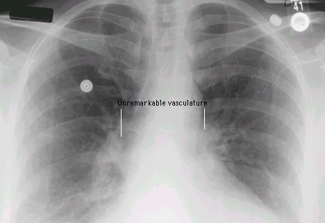 These conditions could be due to an autoimmune disease, a connective tissue disorder, or toxin exposure. Richard B. Gunderman. A Quick Review. To diagnose bibasilar atelectasis, your doctor may order the following tests: CT scan: A chest computed tomography (CT) scan makes precise pictures of your chest structures. Philadelphia, Pa.: Saunders Elsevier; 2016. https://www.clinicalkey.com. Lung nodules are small growths on the lungs. 0. minimal bibasilar atelectasis on ct scan This causes your alveoli to collapse.
Mild Bibasilar Atelectasis in Lungs 1 Mild Bibasilar Atelectasis in Lungs. In medical terms, bibasilar atelectasis meaning describes 2 Causes of Bibasilar Atelectasis. An obstruction to the airflow within the lungs can form in numerous 3 Symptoms of Bibasilar Atelectasis. Bibasilar atelectasis normally just affects a small part The condition is treated based upon what is triggering the condition.