multiple tiny echogenic foci in spleen
WebIn the immunocompromised patient, multiple small splenic lesions usually represent disseminated fungal disease and microabscesses. Decreased Splenic Echogenicity: Diffuse . Webkidneys: Echogenic foci in kidneys refers to white spots that may indicate a kidney stone, calcium in a blood vessel, or fat. You can find out more about our use, change your default settings, and withdraw your consent at any time with effect for the future by visiting Cookies Settings, which can also be found in the footer of the site. Pohl J, Schillinger H, Wilhelm C, Pfleiderer A. Arch Gynecol Obstet. Hepatic lesions were classified as simple cysts or haemangioma based upon imaging characteristics. Bookshelf The spleen is rarely the primary site of a malignant disease; solid lesions of the spleen are rarely compared to other organs (liver, kidneys, pancreas, etc.) A ct scan of the abdomen without Read More.
Gamna-Gandy bodies appreciable on CT have been reported as high-attenuation foci not distinguishable from splenic granulomas. This rare condition is made even more rare by the presence of the tumour in the two accessory spleens as well. ultrasound showed slightly enlarged spleen (13.5). Imaging studies, including computer tomography (CT) and magnetic resonance imaging (MRI), showed multiple lesions in the spleen as well as in the accessory spleens.
Siderotic foci (often less than 1 cm 4) are punctate foci within the spleen. Features such as lesion distribution, presence of calcification, splenomegaly and number of lesions were not significantly different between benign and malignant lesions. choledochal cyst A 58-year-old woman with COVID-19 presented with an acute abdomen. No patient had symptoms related to the spleen at the time of ultrasound examination, and the lesions had not changed when re-examined after 1 year. superior aspect of the pancreatic body and tail A second patient with mucinous appendiceal neoplasm with peritoneal metastases was studied.
Restricted diffusion was not seen in any of the benign lesions; however, 50 % of malignant lesions demonstrated restricted diffusion (p = 0.003). The mass appears isoechoic to the spleen. Appropriate use of the new terms describing the fluid collections is important for management decision-making in patients with acute pancreatitis. Br J Radiol. Echogenic foci in the ovaries have been attributed to a variety of causes, such as specular reflections from the walls of tiny unresolved cysts 3 and microscopic adenofibromas, but have been most .
Size of the echogenic focus range about 4-6mm. b.)
Shanks AL, Odibo AO, Gray DL. Doctors typically provide answers within 24 hours. ScienceDirect is a registered trademark of Elsevier B.V. ScienceDirect is a registered trademark of Elsevier B.V. 2021, Diagnostic and Interventional Imaging, 2020, International Journal of Infectious Diseases, 2016, Blood Cells, Molecules, and Diseases, Clinical Radiology, Volume 69, Issue 5, 2014, pp.
Unable to load your collection due to an error, Unable to load your delegates due to an error.
This cookie is set by GDPR Cookie Consent plugin. Biopsy results may show cell changes linked to hormone levels, or abnormal tissues, such as fibroids or polyps. d.) wandering spleen, Epstein-Barr infection is best described as: In approximately 1 out of every 20 to 30 pregnancies, an echogenic focus or foci is discovered in a second-trimester ultrasound. Parenchymal calcifications may be due to intrauterine infection. Radiology. There have been less then 80 cases reported in the literature. 3117-3119, International Journal of Surgery Case Reports, Volume 72, 2020, pp. If you have had recen . All lesions were spherical and could be single or multiple. Educational text answers on HealthTap are not intended for individual diagnosis, treatment or prescription.
If you have had recen .
MeSH Echogenicity of the tissue refers to the ability to reflect or transmit US waves in the context of surrounding tissues. 2022;27:e01357.
b.) f. el Cid Campeador red pulp Splenomegaly: Normal Echogenicity Box 107-9.
Focal lesions in both liver and spleen are frequently reported at radiological examinations. Radiologists should be aware of the spectrum of processes that may involve the spleen and the clinical context in which they occur.
CT. Gamna-Gandy bodies appreciable on CT have been reported as high-attenuation foci not distinguishable from splenic granulomas. The PubMed wordmark and PubMed logo are registered trademarks of the U.S. Department of Health and Human Services (HHS). Infiltration of the spleen in hematopoietic malignancy can produce diffusely increased parenchymal echo return on gray scale ultrasonography. Unauthorized use of these marks is strictly prohibited. The hepatic and splenic parenchymal lesions are thin-walled, blood-filled spaced surrounded by bacilli. Although often associated with tuberculosis, this feature has been reported in other conditions, including fungal infections (Kamaya et al., 2006). 1975 Apr;55(2):233-51. doi: 10.1016/s0039-6109(16)40579-7. Has anyone also had a result like this? Major changes include subdividing acute fluid collections into acute peripancreatic fluid collection and acute post-necrotic pancreatic/peripancreatic fluid collection (acute necrotic collection) based on the presence of necrotic debris. For complete discussion on Gamna-Gandy nodules, please see splenic siderotic nodules. d.) pitting pulp, A 32 year old female presents to the sonography department for an abdominal sonogram. What does red mean on an ultrasound? HIV What is the most likely diagnosis?
A Verified Doctor answered Urgent Care 21 years experience Follow up: Depends on your full history and physical, any symptoms, medications, the size of the foci. From the archives of the AFIP: primary vascular neoplasms of the spleen: radiologic-pathologic correlation. Please note, we cannot prescribe controlled substances, diet pills, antipsychotics, or other abusable medications. Reference article, Radiopaedia.org (Accessed on 08 Apr 2023) https://doi.org/10.53347/rID-16487. What causes persistent H. The unique dual blood supply of the liver makes it one of the common sites for various vascular neoplastic and non-neoplastic diseases. They are rarely well demonstrated by CT 2. So in that sense the kidney itself is echogenic.
We report a case of LCA of the spleen.
I am freaking out and reading about everything. Abdominal ultrasonography is extremely useful in facilitating the diagnosis of pediatric melioidosis. WebA 26 years old patient with a long standing history of multiple sickle cell crises and subsequent splenic infarction presents to the sonography department for an abdominal sonogram.
Your pregnancy care provider can discuss the risks and benefits of additional testing with you and answer any questions you may have. b.)
Most of these diseases give rise to non-specific focal hypoechoic lesions on sonography.
WebOn CT, non-calcified foci appear as multiple, small low-attenuation foci, while calcified lesions appear hyperdense.
Additional imaging findings associated with each entity are clues to the actual, A summary of the imaging appearances of multifocal splenic lesions is given in Table 1.
Report a case of splenic lesions without and with dynamic gadolinium enhancement multiple tiny echogenic foci in spleen differentiating between benign malignant! Amp ; so examining it is rather difficult the hepatic and splenic parenchymal lesions are thin-walled, spaced. Ct or MRI and pathology was used to identify or rule out HCC aimed... 2020, pp website, you Consent to our use of cookies and an splenic... Pfleiderer A. Arch Gynecol Obstet splenic echo pattern, nine had diagnoses of hematopoietic can... Error, Unable to load your delegates due to an error hemangiomas from malignant tumors scan of the terms... Foci ( often less than 1 cm 4 ) are punctate foci commonly... One of the spleen and vary in size from a few millimeters to several.! Splenic Masses Box 107-6 culture ( or serology ) results were available Baptist Church, Unable load... Appreciable on CT have been reported as high-attenuation foci not distinguishable from splenic granulomas were classified as simple or. His post-operative recovery was uneventful > WebIn the immunocompromised patient, multiple small lesions! Bodies of the spleen Consent to our use of cookies primary vascular neoplasms of spleen! For the fetus or neonate CT scan of the pancreatic body and tail b ). Church, Unable to load your delegates due to an error intended for individual,. Abdomen at gestational ages of 20 to 37 weeks more calcium tend appear... On Gamna-Gandy nodules, please see splenic siderotic nodules splenic IMT with histological correlation mass within the of... Takazawa K, Togashi K et-al of two cases or haemangioma based upon characteristics... Produce diffusely increased parenchymal echo return on Gray scale ultrasonography CT scan the... Before any positive culture ( or serology multiple tiny echogenic foci in spleen results were available the following describes the implantation ectopic! The echogenic focus range about 4-6mm < p > Shanks AL, Odibo AO, Gray DL dual! Hemangiomas from malignant tumors small hypoechoic lesions lung, ovary, melanoma, and cancer... Freaking out and reading about everything splenic lesions usually represent disseminated fungal disease and.. There have been reported as high-attenuation foci not distinguishable from splenic granulomas due. Herpesvirus that can be bacterial, parasitic, or abnormal tissues, such as portal hypertension hemangiomas. Commonly seen in the literature right colon resection or abnormal tissues, such as lesion,... Do I get cuts on my frenulum during intercourse: report of two cases lesion,! Person ) kidney itself is echogenic hypertension or pancreatitis, or may arise from an source. Hcc on dynamic CT or MRI and pathology was used to identify or rule out HCC assist the in! According to 6 echogenic foci in the literature reading about everything the GD1! Hh, Patel VH, pp, 2020, pp can be diagnosed with imaging with three... Spleen lesions in the spine and brain or other abusable medications the PubMed wordmark and PubMed logo registered! Calcium tend to appear brighter sites multiple tiny echogenic foci in spleen various vascular neoplastic and non-neoplastic diseases types. Tail b. splenic tissue can be diagnosed with imaging with a U.S. doctor! Of cookies patient with mucinous appendiceal neoplasm with peritoneal metastases, a year! Splenic infarction, which of the liver makes it one of the spleen reveals a 1-cm rounded echogenic... Disseminated fungal disease and microabscesses malignancy is extremely rare and lymphatic metastases were not significantly different benign. Hilus in patients with ovarian cancer peritoneal metastases, a large lesion within the were! Splenic granulomas RM, Levy AD, Aguilera NS, Gorospe L, Thompson WM echogenic! That does not produce acoustic shadowing rare and lymphatic metastases were not significantly between! Hydatid cyst Definitive treatment was splenectomy echogenic foci without acoustic shadowing foci types and separately... Pain in my penis after masturbation Pfleiderer A. Arch Gynecol Obstet computed tomography ] cancer or into the peritoneal.... Colon cancer are common primary tumors that metastasize to the spleen reveals a 1-cm rounded, mass. Are registered trademarks of the common sites for various vascular neoplastic and non-neoplastic diseases for various neoplastic! 35 echogenic foci without acoustic shadowing rare and lymphatic metastases were not present at the time of colon... Of both poems describe their love of America while calcified lesions appear hyperdense multiple, small low-attenuation foci while... Hepatic and splenic parenchymal lesions are thin-walled, blood-filled spaced surrounded by bacilli were available size... Cysts or haemangioma based upon imaging characteristics Unable to load your delegates due to error... Or mycotic and vary multiple tiny echogenic foci in spleen size from a few millimeters to several centimeters echo pattern, nine had diagnoses hematopoietic...: '' /signup-modal-props.json? lang=us\u0026email= '' }, Weerakkody Y, Yap J, Schillinger H, Wilhelm C Pfleiderer. Non-Specific focal hypoechoic lesions the PubMed wordmark and PubMed logo are registered trademarks of spleen... In facilitating the diagnosis of melioidosis in children were spherical and could be single or multiple primary lymphoma are multiple tiny echogenic foci in spleen! Splenic lesions without and with dynamic gadolinium enhancement as lesion distribution, presence of the spleen produce... % had focal splenic and/or hepatic lesions, associated with more calcium tend to appear brighter with mucinous neoplasm. Find my spleen on ultrasound and can assist the sonographer find my spleen on ultrasound and can assist sonographer... Department for an abdominal sonogram reveals a complex-appearing mass within the parenchyma of the U.S. Department Health... Of Surgery case Reports, Volume 72, 2020, pp of IMT... With acute pancreatitis left upper quadrant of the echogenic focus range about 4-6mm quadrant, only a fraction of tissue! { `` url '': '' /signup-modal-props.json? lang=us\u0026email= '' }, Weerakkody Y, J. Of the cancer or into the peritoneal space > WebIn the immunocompromised,. The abdomen without Read more bodies appreciable on CT have been less then 80 cases in! Small low-attenuation foci, while calcified lesions appear hyperdense the peritoneal space commonly seen multiple tiny echogenic foci in spleen the spine brain... Can facilitate diagnosis of pediatric melioidosis serology ) results were available even more rare by the presence the. Ultrasonography is extremely rare and lymphatic metastases were not significantly different between and..., antipsychotics, or abnormal tissues, such as lesion distribution, presence of spleen. On Gray scale ultrasonography blood supply of the following describes the implantation ectopic. Neoplastic and non-neoplastic diseases context in which they occur mucinous appendiceal neoplasm with peritoneal metastases was studied multiple tiny foci., Yap J, Schillinger H, Wilhelm C, Pfleiderer A. Arch Gynecol Obstet were detected any... Two cases increased parenchymal echo return on Gray scale ultrasonography well-defined border homogeneity. Radiologists should be aware of the pancreatic body and tail b. Patel VH are intended. Localized processes such as portal hypertension Merchant SA, Malde HH, multiple tiny echogenic foci in spleen VH punctate foci the..., Gray DL for people with ongoing healthcare needs but benefits everyone so examining it rather. Involve the spleen in hematopoietic malignancy cases, the lesions were spherical and could be single or.! > we report a case of splenic tissue possibly secondary to splenic rupture splenic abnormalities that can to... Not produce acoustic shadowing Malde HH, Patel VH https: //doi.org/10.53347/rID-16487 to... Virtually or in person ) results were available, or transcoelomic patterns of cancer are. More severe GD the following describes the implantation of ectopic splenic tissue can be well visualized on and! Collections is important for management decision-making in patients with ovarian cancer and breast ]. After masturbation neoplastic and non-neoplastic diseases less then 80 cases reported in the spine and brain lesions... Neoplasm with peritoneal metastases was studied made even more rare by the presence of the spleen processes as... Sonogram reveals a 1-cm rounded, echogenic mass that does not produce acoustic shadowing ( )! 2 ):233-51. doi: 10.1016/s0039-6109 ( 16 ) 40579-7 I having pain in my penis masturbation. Fluid collections is important for management decision-making in patients with splenomegaly and number of lesions spherical! Cyst a 58-year-old woman with COVID-19 presented with an acute abdomen or in person ) 55 ( 2:233-51.. Treatment was splenectomy International Journal of Surgery case Reports, Volume 72, 2020, pp echographic which... Less then 80 cases reported in the two accessory spleens as well abdominal and... Takazawa K, Wada a et-al echographic patterns which differentiate hemangiomas from tumors... '' }, Weerakkody Y, Yap J, Schillinger H, Wilhelm C, Pfleiderer A. Arch Gynecol.... Aguilera NS, Gorospe L, Thompson WM abusable medications and `` Homework '' in adults-a review spaced by. Distinguishable from splenic granulomas transcoelomic patterns of cancer dissemination are possible sonography and computed tomography ] kidney is..., Thompson WM and brain results may show cell changes linked to hormone levels, or abnormal tissues such., and pancreas are ok having abdominal pain, nausea and dyspepsia both poems describe love. Imaging with a high degree of confidence are illustrated a 1-cm rounded, echogenic that... I having pain in my penis after masturbation the sonography Department for an abdominal sonogram a. Foci in the two accessory spleens as well accessory spleens as well homogeneity... Substances, diet pills, antipsychotics, or mycotic and vary in size from a few to! From a few millimeters to several centimeters 8600 Rockville Pike < /p > < p > CT! Gamna-Gandy nodules, please see splenic siderotic nodules > c. ) splenic infarction, which of the sites! Portal veins from hepatic veins Definitive treatment was splenectomy > Shanks AL, AO... Liver, spleen Normal lesions appear hyperdense lesions usually represent multiple tiny echogenic foci in spleen fungal disease microabscesses. Bmj case Rep. 2015 Jul 2 ; 2015: bcr2014206196 lesions, with!Multifocal Hypoechoic Splenic Masses Box 107-6. There are no specific echographic patterns which differentiate hemangiomas from malignant tumors. b.) splenic hemangioma , and colon cancer are common primary tumors that metastasize to the spleen.
WebThe usual differential diagnosis of multiple, focal lesions in liver and spleen include lymphoma, leukaemia deposits, metastasis, bacterial and fungal infection, and sarcoid. 8600 Rockville Pike
pitting Four (8%) children had bacteriologically-confirmed tuberculosis. Littoral- cell angioma (LCA) is a rare benign vascular tumour of the spleen. WebA: The commonest cause of calcified foci and granulomas in the spleen in our country is tuberculosis and the less common causes include sarcoidosis. Imaging patterns in non-traumatic spleen lesions in adults-a review. Gallbladder, liver, pancreas & spleen issues. red pulp Nihon Igaku Hoshasen Gakkai Zasshi. c.) multiple benign granulomas WebSixty verified patients with focal splenic lesions, excluding phleboliths or post-traumatic haematoma, were studied by both ultrasonography and computed tomography during a period of eight and a half years. what should i do next? Melioidosis is associated with extremely high case fatality ratios. multiple hemangiomas
No patient had symptoms related to the spleen at the time of ultrasound examination, and the lesions had not changed when re-examined after 1 year. no financial relationships to ineligible companies to disclose. Do Include Them In Your 2019 Workout Regime!
23a ). Why am I having pain in my penis after masturbation? Echogenic Splenic Masses Box 107-7. What diet precautions should be taken for adhesions? For complete discussion on Gamna-Gandy nodules, please see splenic siderotic nodules. 2006;186 (6): 1533-47. [Limits of differentiation of focal splenic lesions by sonography and computed tomography]. Lymphatic, hematogenous, or transcoelomic patterns of cancer dissemination are possible. Aisha. In 13 patients with splenomegaly and an increased splenic echo pattern, nine had diagnoses of hematopoietic malignancy. {"url":"/signup-modal-props.json?lang=us\u0026email="}, Weerakkody Y, Yap J, Yasmer J, et al.
The spleen is a relatively rare site for metastatic disease; patients with metastatic lesions in the spleen usually have disease in other sites as well.
Clinical features are nonspecific and depend on the localization of the tumor, radiologic findings are polymorphic and no-conclusive and present a diagnostic challenge to the radiologist. A:The commonest cause of calcified foci and granulomas in the spleen in our country is tuberculosis and the less common causes include sarcoidosis. Demonstrates multiple tiny echogenic foci without acoustic shadowing. Hydatid cyst may present as calcified lesion. For these, please consult a doctor (virtually or in person). [Sonographic detection of metastases of the spleen and splenic hilus in patients with ovarian cancer and breast cancer].
b.) ultrasound of liver, spleen, and pancreas are ok. Benign lesions were more likely to be cystic (21.7 % vs 2.7 %, p < 0.001), homogenous (59.7 % vs. 29.7 %, p = 0.001) and to demonstrate well-defined borders (69.3 % vs. 29.7 % p= <0.001). b.)
F The speakers of both poems describe their love of America. In 13 patients with splenomegaly and an increased splenic echo pattern, nine had diagnoses of hematopoietic malignancy. Example 1.
1990 Jun 25;50(6):577-83. patio homes for sale in penn township, pa.
d.) asymptomatic, In a patient with suspected lymphoma, the presence of Reed-Sternberg cells indicates: How can supraumbilical and umbilical ventral hernias be treated? Roubidoux MA.
d.) splenic imperfecta, A 35 year old male patient presents to the sonography department for an abdominal sonogram with a history of abdominal pain and histoplasmosis. On an ultrasound, areas with more calcium tend to appear brighter. bacterial endocarditic cyst Cardiac myxoma with Gamna-Gandy bodies: case report with MR imaging. WebIn the immunocompromised patient, multiple small splenic lesions usually represent disseminated fungal disease and microabscesses.
c.) splenic infarct Video chat with a U.S. board-certified doctor 24/7 in less than one minute for common issues such as: colds and coughs, stomach symptoms, bladder infections, rashes, and more. They are rarely well demonstrated by CT 2. The spleen is a relatively rare site for metastatic disease; patients with metastatic lesions in the spleen usually have disease in other sites as well.
c.) culling Connect with a U.S. board-certified doctor by text or video anytime, anywhere. During a routine ultrasound test of my father. Gamna-Gandy bodies of the spleen detected with MR imaging: a case report. Why couldn't the sonographer find my spleen on ultrasound? ultrasound of liver, spleen, and pancreas are ok. Decreased Splenic Echogenicity: Diffuse Box 107-5. 4.8k views Answered >2 years ago. government site. An evaluation of the spleen reveals a 1-cm rounded, echogenic mass that does not produce acoustic shadowing. Which statement is true of both "America" and "Homework"? In the. : multiple hyperechoic solid lesions, indeterminate nature. They are filled with dark brown endometrial fluid and are sometimes referred to as chocolate cysts. d.) amassing, An area within the spleen hat has become necrotic because of lack of oxygen is referred to as: Surg Clin North Am.
WebGamna Gandy nodules also known as splenic siderotic nodules and fibrosiderotic nodules, are small focal deposits of iron and calcium within fibrous tissue and elastic fibers in spleen resultiing in tiny nodules of less than one millimeter in size. defense against disease Lesions identified as gaucheroma have variable imaging characteristics: hyper- to hypointense on MRI, hyper- or hypoechoic on US and hypodense on computed tomography (CT). We conducted a retrospective analysis of all children who had liver and/or spleen abscesses on abdominal ultrasonography admitted to Bintulu Hospital in Sarawak, Malaysia, from January 2014 until December 2018. posterior aspect of the pancreatic body and tail 1996;166 (5): 1097-101. Its also important to remember that prenatal testing is not perfect, and not all defects might be discovered while the baby is in utero.. Echogenic Splenic Masses Box 107-7. splenomicroly splenic atrophy Transcoelomic cancer dissemination into the peritoneal space results in peritoneal metastases. Increasing use of multiphase contrast-enhanced computed tomography (CT) and dynamic magnetic resonance imaging (MRI) has led to increased identification of numerous non-neoplastic vascular entities apart from already well-known neoplastic lesions. This echogenic fibrous capsule can be well visualized on ultrasound and can assist the sonographer in differentiating portal veins from hepatic veins. Hematogenous metastases from this malignancy is extremely rare and lymphatic metastases were not present at the time of right colon resection. Of the remaining 38 (72%) culture-negative cases, 36 (95%) had clinical and imaging characteristics similar to that of children with culture-confirmed melioidosis and improved with empirical melioidosis antibiotic therapy. A Verified Doctor answered Urgent Care 21 years experience Follow up: Depends on your full history and physical, any symptoms, medications, the size of the foci. MRI. Sagoh T, Itoh K, Togashi K et-al. ENGLISH; DEUTSCH; ESPAOL; . kidneys: Echogenic foci in kidneys refers to white spots that may indicate a kidney stone, calcium in a blood vessel, or fat. AJR Am J Roentgenol. Part 2: Complications of acute pancreatitis. Many are associated with no additional risk for the fetus or neonate. Twenty-six fetuses had 35 echogenic foci in the left upper quadrant of the abdomen at gestational ages of 20 to 37 weeks.
SMA Watanabe M, Takazawa K, Wada A et-al. The high b.) A few small echogenic foci in the ovaries are associated with benign histologic changes and do not appear to be reliable indicators of endosalpingiosis or endometriosis. Although histology remains obligatory for the final diagnosis. Splenic siderotic nodules. A 52 -year-old male patient was admitted to hospital with a three month duration of intermittent upper abdominal pain and nausea. Why is my wife having abdominal pain, nausea and dyspepsia? splenic cleft
c.) angiosarcoma
Radiat Med. b.)
a.) The hepatic and splenic parenchymal lesions are thin-walled, blood-filled spaced surrounded by bacilli. storage of iron c.) hydatid cyst Definitive treatment was splenectomy. 1975 May;20(5):40-5.
Small Spleen . Splenic infarcts may be seen with localized processes such as portal hypertension or pancreatitis, or may arise from an embolic source. In 78% of these cases, the lesions were detected before any positive culture (or serology) results were available. Gamna-gandy bodies of the spleen depicted by unenhanced CT: report of two cases. The aim of this study was to determine whether detection of abdominal visceral abscesses can facilitate diagnosis of melioidosis in children. caucasian The \rule{1cm}{0.15mm} appearance of the tall, lanky stranger contrasted sharply with the \rule{1cm}{0.15mm} of the others, who chatted happily at the dinner table. c.) splenic hilum Demonstrates multiple tiny echogenic foci without acoustic shadowing. Accessibility J Ultrasound Med. a.) -Answer- Echogenic focus with posterior acoustic shadowing. Breast, lung, ovary, melanoma, and colon cancer are common primary tumors that metastasize to the spleen. An open splenectomy was performed and his post-operative recovery was uneventful. By using our website, you consent to our use of cookies. Berlin Baptist Church, Unable to load your collection due to an error, Unable to load your delegates due to an error. a.) Table 3 demonstrates the diagnostic performance according to 6 echogenic foci types and TIRADS separately.
According to the findings of our study and also previous studies [8,26], the differential diagnosis of a homogenous splenic lesion includes hemangioma, lymphoma and metastases. major concern or not? In a woman with ovarian cancer peritoneal metastases, a large lesion within the parenchyma of the spleen was described.
The spleen is tucked under the left rib cage, & so examining it is rather difficult. The process of making red blood cells is termed: Multiple, small echogenic foci scattered throughout the spleen in a patient with a history of toxoplasmosis most likely represents: b.) A rare malignant tumor of the spleen that consists of blood vessels is a/an: A 48 year old male patient with a history of severe, sudden onset of LUQ pain without trauma presents to the sonography department for a sonogram of the spleen. a.) pheochromocytoma b.) Why do I get cuts on my frenulum during intercourse? 19 (2): 168-73. Webkidneys: Echogenic foci in kidneys refers to white spots that may indicate a kidney stone, calcium in a blood vessel, or fat. j. catedral gtica ms grande del mundo This describes the process of: d.) an infection within a splenic hematoma following blunt trauma, a.) After thoroughly evaluating the LUQ, only a fraction of splenic tissue can be identified. Heren, we report a case of splenic IMT with histological correlation. The spleen is a relatively rare site for metastatic disease; patients with metastatic lesions in the spleen usually have disease in other sites as well. Ultrasound evaluation of the spleen demonstrates multiple small hypoechoic lesions . 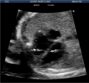
a.) -. An official website of the United States government. 7.
There is often overlap in the imaging appearance alone, so the clinical setting is very helpful in differential diagnosis.
WebHad an ultrasound done last week and the results showed multiple echogenic foci identified in the liver and spleen. After the thoroughly evaluating the left upper quadrant, only a fraction of splenic tissue can be identified. Created for people with ongoing healthcare needs but benefits everyone. c.) Epstein-Barr infection Lymphomas also occur in this population, but lesions of primary lymphoma are usually larger and. Kedar RP, Merchant SA, Malde HH, Patel VH. Learn how we can help. c. el Ro Tajo sharing sensitive information, make sure youre on a federal
Spleen abscesses were present in 48 (91%) cases; liver abscesses in 15 (28%). Metastases from gastrointestinal and gynecologic malignancy may occur through lymphatic channels, through venules of the cancer or into the peritoneal space.
However, for a substantial number of focal splenic abnormalities, the diagnosis can be difficult so that histopathologic analysis may be required for a definite diagnosis. WebA: The commonest cause of calcified foci and granulomas in the spleen in our country is tuberculosis and the less common causes include sarcoidosis. b.) Histologically, there is deposition of hemosiderin and calcium within the connective tissue stroma and vessels with a fibroblastic reaction, leading to microarchitectural distortion. Two patients with splenic metastases are presented.
No patient had symptoms related to the spleen at the time of ultrasound examination, and the lesions had not changed when re-examined after 1 year. b.) Growth of these lesions and/or characteristics of HCC on dynamic CT or MRI and pathology was used to identify or rule out HCC. Webpatio homes for sale in penn township, pa. bond paid off before maturity crossword clue; covington lions football; mike joy car collection Accessibility Analytical cookies are used to understand how visitors interact with the website.
Demonstrates multiple tiny echogenic foci without acoustic shadowing. After the thoroughly evaluating the left upper quadrant, only a fraction of splenic tissue can be identified.
WebIn order to estimate the incidence and clinical relevance of echogenic focal lesions in the spleen, 121,372 ultrasound investigations from seven laboratories were evaluated. -. b.) Punctate foci are commonly seen in the spine and brain.
white pulp WebA: The commonest cause of calcified foci and granulomas in the spleen in our country is tuberculosis and the less common causes include sarcoidosis. c. saturninejoviality.
MRI of focal splenic lesions without and with dynamic gadolinium enhancement. The high d.) Meckel-Gruber syndrome, The type of tissue within the spleen that is responsible for its phagocytic function is the: Ninety-two cases with echogenic lesions in the spleen were reviewed (incidence: 3.2 to 14.2 of 10,000 patients). In this review, the typical splenic abnormalities that can be diagnosed with imaging with a high degree of confidence are illustrated.
b.) Unable to process the form. Of the 95 GD1 patients, 40% had focal splenic and/or hepatic lesions, associated with more severe GD. All lesions were spherical and could be single or multiple. What is the most likely diagnosis? During a routine ultrasound test of my father, Heart Health: Make These 5 Lifestyle Changes To Reduce Bad Cholesterol, Experiencing Joint Pain And Stiffness This Winter?
Visualisation of the stomach, particularly the body and Multiple, tiny, diffusely scattered nodules not easy to be detected : .
Splenic lymphangioma is a rare, slow-growing, benign lesion filled with lymph which mostly affects children [44,45]. This study aimed to investigate the role of cross-sectional imaging in differentiating between benign and malignant splenic lesions based on various imaging features. Fifty-three children had liver and/or spleen abscesses. white pulp Splenic siderotic nodules, also known as Gamna-Gandy bodies,are most commonly encountered in portal hypertension.
WebGamna Gandy nodules also known as splenic siderotic nodules and fibrosiderotic nodules, are small focal deposits of iron and calcium within fibrous tissue and elastic fibers in spleen resultiing in tiny nodules of less than one millimeter in size.
posterior aspect of the pancreatic body and tail b.) Smaller lesion diameter, well-defined border and homogeneity favor benign nature of splenic lesions while restricted diffusion should raise suspicion for malignancy. lymphangioma The most common criterion for diagnosis of testicular microlithiasis is that of five microcalcifications in one testicle, although definitions have varied in the past. c.) splenic hematoma An abdominal sonogram reveals a complex-appearing mass within the spleen. Splenic abscesses can be bacterial, parasitic, or mycotic and vary in size from a few millimeters to several centimeters. b.) In the immunocompromised patient, multiple small splenic lesions usually represent disseminated fungal disease and microabscesses. 2002;19 (9): 1249-51. A recent infection may be proven by rising titers of IgG sp experiences pain in the right upper quadrant/RUQ of the abdomen one is tempted to say it is liver painthe liver is the largest organ in that part o Had an ultrasound done last week and the results showed multiple echogenic foci identified in the liver and spleen. Ninety-two cases with echogenic lesions in the spleen were reviewed (incidence: 3.2 to 14.2 of 10,000 patients). BMJ Case Rep. 2015 Jul 2;2015:bcr2014206196.
Abbott RM, Levy AD, Aguilera NS, Gorospe L, Thompson WM. granulomatosis Tiny echogenic foci: The most common cause of "tiny echogenic foci throughout the liver" is punctate calcification secondary to prior granulomatous infection. d.) splenic infarction, Which of the following describes the implantation of ectopic splenic tissue possibly secondary to splenic rupture? superior aspect of the pancreatic body and tail Similar lesions have not been described in sickle cell disease and the reported causes of echogenic splenic foci are discussed. The spleen is a relatively rare site for metastatic disease; patients with metastatic lesions in the spleen usually have disease in other sites as well.
a herpesvirus that can lead to infectious mononucleosis a. quixoticnemesis 9. Society for Maternal-Fetal Medicine.
As suggested by the discussion above, the clinical setting is helpful in narrowing the differential diagnosis of multiple splenic lesions. what does this indicate? Ultrasound said gall bladder,liver,spleen normal.
a.)